

Source: Supra-Gingival Minimally Invasive Dentistry: A Healthier Approach to Esthetic Restorations, published by Wiley-Blackwell
What is Supragingival Minimally Invasive Adhesive Dentistry?
Supra-gingival minimally invasive adhesive dentistry is a method of restorative dentistry that uses a number of modern techniques, as well as the benefits of adhesive dentistry, to avoid crowns or any restorative procedure that may cause preventable subgingival margins.
It is a healthier form of restorative dentistry, as it strives to retain supra-gingival restorative margins and to preserve as much tooth as possible. Preparation, impressions, and cementation are easier, faster, and also more predictable with supra-gingival margins. When subgingival caries or margins are already present, it is also possible to limit and repair the damage with these techniques.
Why Supra-Gingival Minimally Invasive Adhesive Dentistry is Valuable
Supra-gingival minimally invasive adhesive dentistry offers numerous benefits. Although the desire to stay supra-gingival in dentistry is a universal one, the difference is the systematic approach, the many specialized techniques, and the absolute commitment to supra-gingival minimally invasive restorations. Supra-gingival margins in fact make restorative dentistry:
- Easier for the clinical
- Healthier for the patient
- Aesthetically pleasing
- Provide for long lasting, predictable results (Figures 1a-g, 2a,b).

Figure 1
In Figure 1, we see a damaged tooth needing an indirect restoration (a), undisturbed gingiva during onlay preparation and showing very little tooth removal (b), healthy gingival around finished onlay (c), and badly damaged teeth (d).
Then, after the preparation, we see absolutely minimum tooth reduction (e) followed by a cemented restoration (f), with an occlusal view (g).

Figure 2
Then in Figure 2, we see a supra-gingival veneer preparation of a fractured tooth and adjacent central (a) followed by a healthy undisturbed gingiva around the finished veneers (b).
The History
An increased use of new tooth-colored materials followed esthetic demands from patients. Not considered possible before, adhesives have allowed the clinician to provide minimally invasive esthetic dentistry [1,2,3,4,5]. Though, when initially introduced, bonded restorations posed challenges for dental professionals. They were seemingly less predictable and filled with complications, including postoperative sensitivity and restoration fractures, which resulted in their limited use.
A lack of knowledge and a lack of trust in adhesion resulted in a less than optimal use of these new tooth-colored materials. Rather than using tooth-conserving adhesion techniques, traditional mechanically retained techniques were utilized, often proven to be counterproductive.
However, newer materials, improved restorative and adhesion techniques, and the realization of previously hidden advantages, particularly supra-gingival margins [6,7,8,9], have allowed these restorations to live up to their full potential [10,11,12]. Adhesion facilitates the preservation of healthy tooth structure. Because remaining dentin thickness correlates with continued tooth vitality [13], bonded restorations preserve the vitality of the tooth (Figure 3a-c).

Figure 3
In Figure 3, we see a tooth that may have been treated with a crown (a), then after supra-gingival minimally invasive preparation (b), and finally that same tooth 11 years postoperatively (c).
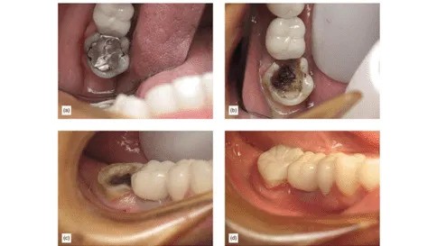
While partial coverage bonded restorations, such as onlays and veneers, are used in dentistry, they are highly under-utilized. The overwhelming majority of clinicians consider them to be a limited alternative to full crowns, to be used only when a number of requirements are fulfilled. [14,15,16,17,18]. However, we see in Figure 4 an extremely damaged tooth with amalgam (a) then the amalgam removed, and if axial reduction was performed the tooth would end up deeply subgingival (b). In image c, we see a preparation that is supra-gingival and easy to isolate an event on a second molar, followed by the finished restoration supported by adhesion (d).

Figure 4
Keeping restorative margins supra-gingival has been left to chance. In the cases where it is not easily achievable, full coverage becomes the preferred choice. Until now, there has not been a specific protocol or set of techniques that have been advocated to prevent subgingival margins.
Advancements in adhesive dentistry, coupled with the great value placed on the health of the periodontium, make supra-gingival margins a priority. Minimally invasive partial coverage restorations, whether direct or indirect, are the preferred option in all cases except for teeth already cut for crowns.
Although traditional mechanically retained restorations may seem more predictable and easier due to their longer history, supra-gingival minimally invasive adhesive dentistry in fact offers a superior restorative alternative for both patients and dentists.
Future articles will consider principles and techniques for supra-gingival dentistry as well as a supra-gingival protocol for achieving supra-gingival margins.
We hope this article was valuable to you. Our mission is to provide proven, real-world practical techniques, resources, articles and videos to help the community of caring dentists, who value the benefits of minimally invasive Supra-gingival dentistry, expand their knowledge and achieve clinical success, thus giving their patients a healthier form of dentistry.
For updates on newly published articles, courses, and more, sign up to the Ruiz Dental Seminars newsletter.
Disclaimer: Los Angeles Institute of Clinical Dentistry & Ruiz Dental Seminars Inc. uses reasonable care in selecting and providing content that is both useful and accurate. Ruiz Dental Seminars is not responsible for any damages or other liabilities (including attorney’s fees) resulting or claimed to result in whole or in part, from actual or alleged problems arising out of the use of this presentation. The techniques, procedures and theories on this presentation are intended to be suggestions only. Any dental professional viewing this presentation must make his or her own decisions about specific treatment for patients.
Sources
- Buonocore MG. A simple method of increasing the adhesion of acrylic filling materials to enamel surfaces. J Dent Res, 1955; 34(6): 849-853.
- Fusayama T, Nakamura M, Kurosaki N, Iwaku M. Non-pressure adhesion of a new adhesive restorative resin. J Dent Res, 1979; 58(4): 1364-1370.
- Bertolotti RL. Total etch: The rational dentin bonding protocol. J Esthet Dent, 1991; 3(1):1-6.
- Calamia JR. The etched porcelain veneer technique. NY State Dent J, 1988; 54(7):48-50.
- Bertolotti RL. Adhesion to porcelain and metal. Dent Clin North Am, 2007; 51(2):433-51, ix-x.
- Ruiz JL. Simplifying the cementation of porcelain onlays. Dent Today, 2004; 23(3): 76-79.
- Ruiz JL. Supragingival dentistry using metal-free restoration. Dent Today, 2008; 27: 104-109.
- Ruiz JL. Anterior and posterior partial-coverage indirect restorations using supragingival dentistry techniques. J Mass Dent Soc, 2012; 61(2): 16-19.
- Ruiz JL, Kurtz R. Are full crowns over utilized? Supragingival partial-coverage designs as a first option. Dent Today, 2014; 33(5): 124-125.
- Magne P, Douglas WH. Rationalization of esthetic restorative dentistry based on biomimetics. J Esthet Dent, 1999; 11(1): 5-15.
- Kramer N, Frankenberger R. Clinical performance of bonded leucite-reinforced glass ceramic inlays and onlays after 8 years. Dent Mater, 2005; 21: 267-271.
- Posselt A, Kerschbaum T. Longevity of 2328 chairside CEREC inlays and onlays. Int J Comput Dent, 200; 6(3): 231-248.
- Zollner A, Gaengler P. Pulp reactions to different preparation techniques on teeth exhibiting periodontal disease. J Oral Rehabil, 2000; 27(2): 93-102.
- Bakeman EM, Kois JC. Posterior, all-porcelain, adhesively retained restoration. Inside Dentistry, 2009; 5(5): 20-30.
- Kois DE, Chaiyabutr Y, Kois JC. Comparison of load fatigue performance of posterior ceramic onlay restorations under different preparation designs. Compend Contin Educ Dent, 2012; 33 (Spec No 2): 2-9.
- Meye A Jr, Cardoso LC, Araujo E, Baratieri LN. Ceramic inlays and onlays: clinical procedures for predictable results. J Esthet Restor Dent, 2003; 15(6): 338-352.
- Barghi N, Berry TG. Clinical evaluation of etched porcelain onlays: A 4-year report. Compend Contin Educ Dent, 2002; 23(7): 657-674.
- Heymann, HO, Swift EJ, Ritter AV. Indirect tooth color restorations, in Sturdevant’s Art and Science of Operative Dentistry, 6th ed. Elsevier Mosby, 2013, pp. 280-295.