

Source: Supra-Gingival Minimally Invasive Dentistry: A Healthier Approach to Esthetic Restorations, published by Wiley-Blackwell
Mechanically Retained Dentistry With Subgingival Margins Is Less Healthy
As already stated, the negative effects of subgingival margins are not usually considered in dentistry. Traditional mechanically retained dentistry uses multiple techniques and protocols which leave subgingival margins. But what are the consequences of these techniques? A subgingival margin will undoubtedly be a factor that will enhance subgingival plaque accumulation. Waerhaug showed, in research with extracted human teeth, that 9 of 10 restorations with subgingival margins were covered with plaque. The conclusion was that “restorations placed below the gingival margin indirectly are strongly involved in the etiology of destructive periodontal disease” (Figure 1) [1].
Of course, the worse the margin adaptation, the worse the plaque accumulation. Poor margins lead to what I refer to as “permanent tartar.” Permanent tartar is the overhang that traps bacteria and food, becoming destructive to the health of the gingiva (Figure 2). It is difficult to see and remove excess cement when it is hidden below the gum line. In another human study, Muller showed that the location of the margin (supragingival, equigingival, or subgingival) clearly affects periodontal health [2]. Larato, in a clinical study, found a difference of almost 1 mm in pocket depth between non-restored teeth and teeth with crowns with subgingival margin placement [3]. Beyond research, clinical experience clearly shows the severed damage that subgingival margins cause to the periodontium (Figures 3, 4). In addition, techniques such as cord packing used with traditional subgingival margin placement have the potential to be detrimental to periodontal health [4,5], especially when performed aggressively and incorrectly (Figure 5).

Figure 1: A dentist who had some prior subgingival veneers done, clearly showing inflammation on veneers and healthy gums elsewhere.

Figure 2: Severe gingival inflammation showing the effects of poor margin fit and accumulation of debris and tartar.

Figure 3: Observe the severe inflammation on tooth #9 caused by deep subgingival margin.

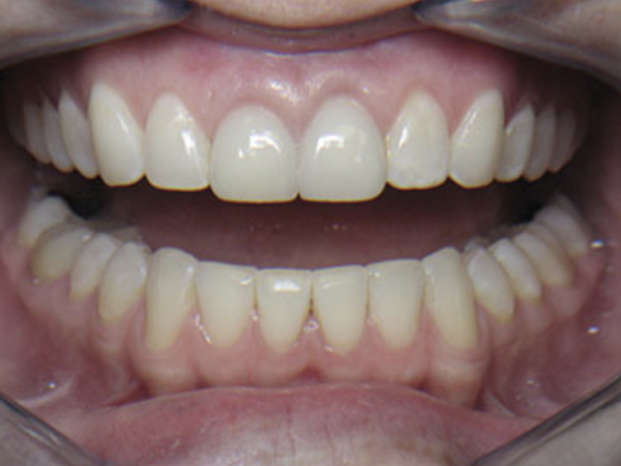
Figure 4: Observe the healthy periodontal condition of all non-crowned teeth.

Figure 5: Aggressive cord must be placed with deep subgingival margins, causing much irritation.
Additionally, the more subgingival the restorative margin, the more tooth is reduced. As demonstrated by Shillingburg, “the effect of apical reduction” applies. This means that the more apically the margin is placed, the closer to the pup the preparation becomes, since a taper in the crown preparation must be maintained (Figure 6) [6]. The effects of axial reduction and other mechanical features on the pulpal health are considerable.

Figure 6: The effect of apical reduction.
Supragingival Minimally Invasive Adhesive Dentistry Is Healthier
Not having to compromise or invade the periodontium is one of the chief reasons that supragingival dentistry is healthier for our patients. Poor gingival health is often seen surrounding porcelain-fused-to-metal (PFM) crowns, while supragingival restorations do not affect periodontal health (Figure 7). The relationship between subgingival margins and poor periodontal health cannot be overemphasized [7,8,9].
Gingival recession is also less likely to occur when gums are undamaged by subgingival preparation and aggressive cord packing. Supragingival restorative techniques eliminate these problems (Figure 8a,b). Even in cases when the clinician may not be able to achieve a perfect marginal fit and smoothness, when the margin is supragingival, the consequences of the imperfect margins are not as devastating to the periodontium. Supragingival defective margins will be more accessible for patients to clean and maintain, and the clinician will be able, at a later, to realize and perhaps repair the defect because of its better visibility both on x-ray and visually. All of the above provides patients with better quality, healthier dentistry.

Figure 7: Compare periodontal health around the onlay on the lower second molar and the subgingival margin maxillary crown.

Figure 8: (a) Healthy gingiva on onlays with visible supragingival margins. (b) Healthy gingiva on veneers with visible supragingival margins.
Pulpal health is another major benefit of supragingival adhesive dentistry. FIrst, avoiding the need for mechanical retention with the health of adhesion avoids unnecessarily removal of millimeters of healthy tooth and thus overheating and traumatizing the pulp (Figure 9a,b). Supragingival bonded dentistry allows the dentist to work away from vital pulp because it does not require axial reduction or any other mechanical retention features. Proximity to the pulp during preparation has been clearly shown to increase both pulp temperature and pulpal changes [10,11,12,13]. Innumerable root canals can be avoided by remaining far away from the nerve during full crown preparation [14].

Figure 9: Comparison of (a) onlay preparation with only occlusal reduction and (b) crown preparation getting closer to the pulp.
Finally, the value of preserving natural, healthy tooth structure that can be achieved with supragingival minimally invasive adhesive dentistry cannot be stressed enough [15]. The strength of natural, healthy enamel and dentin will always be greater than any other restorative material (Figure 10).

Figure 10: My standard tooth reduction comparison: virgin, onlay, crown.
We hope this article was valuable to you. Our mission is to provide proven, real-world practical techniques, resources, articles and videos to help the community of caring dentists, who value the benefits of minimally invasive Supra-gingival dentistry, expand their knowledge and achieve clinical success, thus giving their patients a healthier form of dentistry.
For updates on newly published articles, courses, and more, sign up to the Ruiz Dental Seminars newsletter.
Disclaimer: Los Angeles Institute of Clinical Dentistry & Ruiz Dental Seminars Inc. uses reasonable care in selecting and providing content that is both useful and accurate. Ruiz Dental Seminars is not responsible for any damages or other liabilities (including attorney’s fees) resulting or claimed to result in whole or in part, from actual or alleged problems arising out of the use of this presentation. The techniques, procedures and theories on this presentation are intended to be suggestions only. Any dental professional viewing this presentation must make his or her own decisions about specific treatment for patients.
Sources
- Waerhaug J. Presence or absence of plaque on subgingival restorations. Scand J Dent Res, 1975; 83(1): 193-201.
- Müller HP. The effect of artificial crown margins at the gingival margin on the periodontal conditions in a group of periodontally supervised patients treated with fixed bridges. J Clin Periodontal, 1986; 13(2): 97-102.
- Larato DC. Effects of artificial crown margin extension and tooth brushing frequency on gingival pocket depth. J Prosthet Dent, 1975; 34(6): 640-643.
- Polat NT, Ozdemir AK, Turgut M. Effects of gingival retraction materials on gingival blood flow. Int J Prosthodont, 2007; 20(1): 57-62.
- Fazekas A, Csempesz F, Csabai Z, Vág J. Effects of presoaked retraction cords on the microcirculation of the human gingival margin. Oper Dent, 2002; 27(4): 343-348.
- Shillingburg HT, Hobo S, Whitsett LD, Jacobi R, Bracket SE (eds). Principles of tooth preparations, in Fundamentals of Fixed Prosthodontics, 3rd ed. Quintessence Books; 1997, pp. 199-135.
- Larato DC. Effect of cervical margins on gingiva. J Calif Dent Assoc, 1969; 45: 19-22.
- Silness J. Periodontal conditions in patients treated with dental bridges. J Periodontal Res, 1970; 5: 6-68.
- Williams DF, Smith DC. Biocompatibility of Dental Materials. Boca Raton, FL: CRC Press; 1982.
- Thomas MS, Kundabala M. Pulp hyperthermia during tooth preparation: The effect of rotatory instruments, laser, ultrasonic devices and airborne particle abrasion. J Calif Dent Assoc, 2012; 40(9): 721-731.
- Davis GR, Tayeb RA, Seymour KG, Cherukara GP. Quantification of residual dentine thickness following crown preparation. J Dent, 2012; 40(7): 571-576.
- Langeland K, Langeland LK. Pulp reactions to cavity and crown preparation. Aust Dent J, 1970; 5(4): 261-276.
- Dahl BL. Dentine/pulp reactions to full crown preparation procedures. Oral Rehabil, 1977; 4(3): 247-254.
- Valderhaug J, Jokstad A, Ambjørnsen E, Norheim PW. Assessment of the periapical and clinical status of crowned teeth over 25 years. J Dent, 1997; 25: 97-105.
- Ruiz JL. Supragingival dentistry using metal-free restorations. Dent Today, 2008; 27: 104-109.