

Source: Supra-Gingival Minimally Invasive Dentistry: A Healthier Approach to Esthetic Restorations, published by Wiley-Blackwell
The choice of restorative materials for partial or full coverage supragingival minimally invasive onlays or partial crowns is of paramount importance. Patient satisfaction and the success and longevity of the restoration depend on this choice. There are many restorative materials for partial coverage onlays, including gold.
Bertolotti has outlined the techniques for bonding gold to a tooth [1]. Currently, gold has diminished in popularity because it is not esthetic and is expensive. However, it should be considered for non-visible short teeth such as maxillary second molars. Gold does not fracture, so the restoration can be made thinner than ceramics, which is ideal for short teeth. Bonded partial coverage onlays or overlays can be made of different types of non-metal materials: Laboratory composite, leucite-reinforced pressed feldspathic porcelain, lithium, disilicate and zirconia. Bonding with zirconia has been questioned, probably because it cannot be etched with hydrofluoric acid like most other ceramics. However, with the appropriate technique, zirconia can be bonded reasonably well [2,3]. Although not currently a popular material, alumina (aluminum oxide, Al203) can be bonded using the same methods as for zirconia.
Composite
Composite material can be divided into intraoral composite used for semi-direct onlays or laboratory composite used for indirect onlays and computer-aided design and milled (CAD-CAM) composite. The benefits of composite onlays are that they can be easy to fabricate, intrinsically polychromatic, highly esthetic, initially, less expensive, and easy to repair.
Although they may lose luster and look dull over time (Figure 1), composite onlays show good fracture strength, similar to strong ceramic [4]. The disadvantage of composite is its lack of rigidity, which may lead to an increased risk of bond failure and possibly shorter durability [20,5]. Both clinical experience and research indicate that composite wears faster than tooth over the long term [6]. Super-thin composite occlusal veneers, while very strong in relation to fracture, will have a shorter clinical life because of the poor wear resistance of composite (Figure 2a,b) [7]. New hybrid nanocomposites, such as Lava Ultimate (no longer indicated for crowns by 3M but still suitable for bonded onlays) and Z Nano, which combine ceramics and silica nanoparticles, have been shown to be very strong, but only time will tell whether they wear as or better than more conventional composites.

Figure 1: Dull belleGlass composite onlay.

Figure 2: (a) Ten-year onlay recall, both belleGlass onlays damaged owing to excessive composite wear. (b) Nine-year recall showing excessive wear and necessity for replacing a composite onlay due to wear.
Leucite-Reinforced Pressed Feldspathic Porcelain
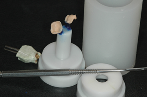
Leucite-reinforced pressed feldspathic porcelain can be layered, pressed (Figure 3) or milled in a CAD-CAM machine. Leucite-reinforced feldspathic pressed restorations have a long history of success for bonded onlays. Millions of successful restorations have been done with these materials and the literature is replete with the excellent results achieved with [8,9]. This material has the capacity to provide the best esthetics and blending of the restorative margin and the tooth because it has ideal translucency. When bonded to a rigid tooth structure, it can be as strong as lithium disilicate in compressive strength [6]. Its shear strength to avoid lateral interferences, which can cause fractures. The primary form of failure for restorations made from this material is bulk fracture (Figure 4a,b) [10,11]. Nevertheless, with sufficient remaining tooth structure, proper adhesion and good attention to occlusion, these materials have performed very well, and I have placed several thousands with overwhelming success (Figure 5).

Figure 3: Waxed onlays being invested.

Figure 4: (a) Many bulk fractures pressabel onlay are related to adhesive failure; the restoration came clean off the tooth. (b) Bulk fracture of pressable onlay, also showing a clean separation of the restoration, showing that this was an adhesive failure.

Figure 5: Three 14-year-old VenusCeram pressed onlays in good condition.
Lithium disilicate
Lithium disilicate can be pressed or milled in a CAD-CAM machine. For multiple reasons, this material has quickly become the most ideal for bonded onlays. When strength is the priority [12,13], the pressed version appears to be a little stronger and provides a better fit [14]. Lithium disilicate strength can exceed 500 MPa, and is much stronger when bonded than when cemented (Figure 6).
When largely bonded to rigid tooth structure such as enamel, its strength can be close to that of zirconia. However, when bonded to a less rigid structure such as dentin, it becomes considerably weaker than zirconia [15]. There are two versions: high and low translucency. High translucency is preferable for most cases of partial coverage onlays where the cavosurface margin is considerably coronal to the gingival line and good blending is important (Figure 7a-c). Low translucency may be preferable if the color of the tooth needs to be hidden, but the disadvantage is that the union of the tooth and restoration will be very visible and will often need to be placed equi- or subgingivally (Figure 8). There are techniques that will allow the color to be corrected first, thus allowing supragingival techniques to be used. As with anything in life, exceptions exist when no other option will be available other than a subgingival margin.

Figure 6: Ivoclar graph of strength of bond compared with cement.

Figure 7: (a) Preparation with bevel for e-Max onlay. (b) E-Max onlay on cast showing area of translucency. (c) Good blending with high translucency e-Max.

Figure 8: Opacious low translucency e-Max will not allow good blending of restorative margins.
Zirconia
Zirconia has a long history of success in dentistry [16,17,18]. Its intrinsic strength is remarkable, exceeding 1,000 MPa. It can be milled to a thin coping, then layered or pressed veneer ceramics can cover the coping, creating a layered restoration, porcelain fused to zirconia (PFZ), similar to the porcelain fused to zirconia (PFZ), similar to the porcelain fused to metal of old. It is opacious (Figure 9) unless very thin. Zirconia can also be made monolithic; that is, a full-contour zirconia without a veneer. This material is more ideally indicated for full crowns and bridge frameworks. Zirconia onlays are usually monolithic and are indicated in cases with lower esthetic requirements, in cases with a high risk of fracture, and in teeth which have lost their rigidity, such as endodontically treated teeth, especially first and second molars. For most onlays, the monolithic option is usually more desirable, as it requires less reduction of the tooth. Because of its poor translucency and lack of ability to blend with the tooth, zirconia onlay preparations require the extension of the restorative margin closer to the gingiva, but ideally not subgingivally. Correct adhesive to zirconia is not as well known, but excellent results can be achieved using a bonding enhancement air abrasive powder [19] (see Chapter 9 for details of adhesion techniques).

Figure 9: Very ugly zirconia margin.
We hope this article was valuable to you. Our mission is to provide proven, real-world practical techniques, resources, articles and videos to help the community of caring dentists, who value the benefits of minimally invasive Supra-gingival dentistry, expand their knowledge and achieve clinical success, thus giving their patients a healthier form of dentistry.
For updates on newly published articles, courses, and more, sign up to the Ruiz Dental Seminars newsletter.
Disclaimer: Los Angeles Institute of Clinical Dentistry & Ruiz Dental Seminars Inc. uses reasonable care in selecting and providing content that is both useful and accurate. Ruiz Dental Seminars is not responsible for any damages or other liabilities (including attorney’s fees) resulting or claimed to result in whole or in part, from actual or alleged problems arising out of the use of this presentation. The techniques, procedures and theories on this presentation are intended to be suggestions only. Any dental professional viewing this presentation must make his or her own decisions about specific treatment for patients.
Sources
- Bertolotti RL. Adhesion to porcelain and metal. Dent Clinics North Am, 2007; 51: 433-451.
- Kern M. Bonding to oxide ceramics: Laboratory testing versus clinical outcome. Dent Mater, 2015; 31(1): 8-14.
- Yang B, Wolfart S, Scharnberg M, Ludwig K, Adelung R, Kern M. Influence of contamination on zirconia ceramic bonding. J Dent Res, 2007; 86(8): 749-753.
- Belli, R. Mechanical fatigue degradation of ceramics versus resin composites for dental restorations. Dent Mater, 2014; 30(4): 424-432.
- Hopp C, Land MF. Considerations for ceramic inlays in posterior teeth: a review. Clin Cosmet Investig Dent, 2013; 5:21-32.
- Yesil ZD, Alapati S, Johnston W, Seghi RR. Evaluation of the wear resistance of new nanocomposite resin restorative materials. J Prosthet Dent 2008; 99(6): 435-443.
- Magne P, Stanley K, Schlichting LH. Modeling of ultrathin occlusal veneers. Dent Mater, 2012; 28(7): 777-782.
- Frankenberger R, Taschner M, Garcia-Godoy F, Petschelt A, Krämer N. Leucite-reinforced glass ceramic inlays and onlays after 12 years. J Adhes Dent, 2008; 10(5): 393-398.
- Ruiz JL, Christensen GJ, Sameni A, Vargas L. Clinical performance of bonded ceramic and resin-based composite inlays and onlays using a self-etch bonding system; a 51-month report. Inside Dentistry, 2007; 3(5): 62-65.
- El-Mowafy O, Brochu JF. Longevity and clinical performance of IPS-Empress ceramic restorations: a literature review. J Can Dent Assoc, 2002; 68: 233-237.
- Lehner C, Studer S, Brodbeck U, Schärer P. Six-year clinical results of leucite-reinforced glass ceramic inlays and onlays. Acta Med Dent Helv, 1998; 3:137-146.
- Culp L, McLaren EA. Lithium disilicate: the restorative material of multiple options. Compend Contin Educ Dent, 2010; 31(9): 716-725.
- Fasbiner DJ, Dennison JB, Heys D, Neiva G. A clinical evaluation of chairside lithium disilicate CAD/CAM crowns: A two year report. J Am Dent Assoc, 2010; 141(6 Suppl): 10s-14s.
- Anadioti E, Aquilino SA, Gratton DG, Holloway JA, Denry IL, Thomas GW, Qian F. Internal fit of pressed and computer-aided design/computer aided manufacturing ceramic crowns made from digital and conventional impressions. J Prosthet Dent, 2015; 113(4): 304-309.
- Ma, L. Load-bearing properties of minimal-invasive monolithic lithium disilicate and zirconia occlusal onlays: finite element and theoretical analyses. Dent Mater, 2013; 29(7): 742-751.
- Christensen RP, Ploeger BJ, A clinical comparison of zirconia, metal and alumina fixed-prosthesis frameworks veneered with layered or pressed ceramic: a 3 year report. J Am Dent Assoc, 2010; 141(11); 1317-1329.
- Keough BE, Kay HB, Sage RD, Keen E. Clinical performance of scientifically designed, hot isostatic-pressed (HIP’d) zirconia cores in a bilayered all-ceramic system. Compend Contin Educ Dent, 2011; 32(6): 58-68.
- Albashaireh ZS, Ghazal M, Kern M. Two body wear of different ceramic materials opposed to zirconia ceramics. J Prosthet Dent, 2010; 104(2): 105-113.
- Wolfart M, Lehmann F, Wolfart S, Kern M. Durability of the resin bond strength to zirconia ceramic after using different surface conditioning methods. Dent Mater, 2007; 23(1): 45-50.
- Bertolotti RL. Adhesion to porcelain and metal. Dent Clinics North Am, 2007; 51: 433-451.