

Source: Supra-Gingival Minimally Invasive Dentistry: A Healthier Approach to Esthetic Restorations, published by Wiley-Blackwell
Mechanically Retained Dentistry Is Less Beautiful
It is interesting that one of the primary reasons given for placing margins subgingivally in dentistry fails so often. Historically, restorative margins have been placed subgingivally because of the desire to hide the unsightly margin of opacious crowns, usually PFM and, more currently zirconia and IPS e-max (Ivoclar). Subgingival margins are also placed when the color of the tooth is to be changed by the restorative materials, in cases of dark teeth, or endodontically stained teeth. Unfortunately, this technique seldom works well (Figure 1). First, in the short term, opacious materials are usually unnatural and not attractive; the typical “Chiclet” crown is rarely esthetically pleasing (Figures 2 a,b, 3). Teeth have natural translucency, but when they are covered with an opacious restorative layer unnatural things happen. One effect is that the cervical area of the tooth appears dark because the opacious restoration blocks the light from entering the tooth. The lack of light inside the tooth makes it appear darker or more gray, so the blending of the restoration becomes less predictable. This is the primary reason for the dreaded gray gum around crowns (Figure 4). Unhealthy red gingiva is never beautiful.

Figure 1: Very dark root canal treated tooth with opacious crown, with unesthetic results.

Figure 2: (a) Root canal treated tooth restored with opacious crown, leaving a gray margin and “Chiclet” -like tooth. (b) Close-up view of the same tooth; this crown has been replaced three times and each time it ends up showing the margin again.

Figure 3: This patient was very unhappy with the grayness of the gums on all her crowns.

Figure 4: This patient complained that the margin used to be acceptable, but the gums have receded and now she does not like the appearance and wants the crowns replaced.
In the long term, aging and periodontal conditions lead to gingival recession, accelerated by the unhealthy periodontium caused by subgingival margins. When the unsightly margin becomes visible, it often leads to early removal and replacement of the restoration (Figures 4, 5a,b). Proper use of translucent restorations will render this unnecessary, which is discussed in later chapters.

Figure 5: (a) These are clinically healthy crowns, but the patient wanted them replaced. (b) After crown replacement – an extremely difficult case, and most likely the gingiva will recede in a few years.
Supragingival Minimally Invasive Adhesive Dentistry Is More Esthetic
Unlike the traditional approach of hiding the restorative margins subgingivally, supragingival dentistry principles allow for more predictable esthetic results. The correct use of translucent restorative materials to achieve good margin blending is one of the five rules of supragingival restorative dentistry. Translucency is one of the most important aspects of esthetic in dentistry. Teeth have natural translucency, so when translucent material is used correctly, an outcome close to natural can be achieved, resulting in more beautiful natural restorations. Opacious restorations have the opposite effect (Figure 6a,b, 7a,b).
Translucency allows for the ideal blending of tooth and restoration, owing to light passing through the restoration and into the tooth. Enamel and dentin function similarly to fiber optic rods. When light hits the crown, it travels up through the root [1]. Conversely, when light does not pass through the tooth, the root darkens and appears gray (Figure 8).

Figure 6: (a) Translucent natural-looking veneers. (b) Opacious fake-looking anterior crowns.

Figure 7: (a) Compare the enhanced esthetics of the translucent onlay on the second bicuspid with the opacious molar crowns. (b) Opacious fake-looking anterior crown.

Figure 8: (a) Observe how dark the root looks with the opacious crown. (b) Dramatic difference in color after the opacious porcelain-fused-to-metal crown is replaced with translucent ceramic.
PFM, full zirconia, layered zirconia, lithium disilicate, composite, feldspathic pressed and layered range from fully opacious to very translucent [2]. Proper use of translucency is key to supragingival dentistry because, when translucent materials are used, the restoration margin will blend with the tooth, creating something of a “contact lens” effect, ensuring that the margin blends neatly. In the esthetic zone, with higher translucency, the margin will virtually disappear (Figure 9) [3]. Proper use of translucency while choosing the restorative and luting materials allows for the margin placement to be slightly above the gingiva, even in the esthetic zone, and supragingival in the areas with lesser esthetic requirements. Very beautiful results can be achieved in both posterior and anterior regions when the correct materials are used appropriately, which is explained in subsequent chapters.

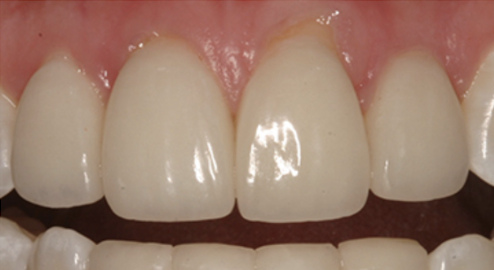
Figure 9: Observe that by using translucent porcelain, the restorative margin can blend to an almost undetectable level, similar to the “contact lens” effect.
Beautiful gums or gingiva are also part of good esthetics. Supragingival dentistry allows for a more beautiful gingival health. Subgingival margins often lead to slightly or even severely inflamed gingiva, a look which is far from beautiful (Figure 10). Supragingival margins allow for perfectly undisturbed, healthy gingiva, which always enhances the final results.

Figure 10: Even well-shaped porcelain restorations look bad with ugly gums.
We hope this article was valuable to you. Our mission is to provide proven, real-world practical techniques, resources, articles and videos to help the community of caring dentists, who value the benefits of minimally invasive Supra-gingival dentistry, expand their knowledge and achieve clinical success, thus giving their patients a healthier form of dentistry.
For updates on newly published articles, courses, and more, sign up to the Ruiz Dental Seminars newsletter and to view more of Dr. Ruiz’s excellent work, check out his smile gallery.
Disclaimer: Los Angeles Institute of Clinical Dentistry & Ruiz Dental Seminars Inc. uses reasonable care in selecting and providing content that is both useful and accurate. Ruiz Dental Seminars is not responsible for any damages or other liabilities (including attorney’s fees) resulting or claimed to result in whole or in part, from actual or alleged problems arising out of the use of this presentation. The techniques, procedures and theories on this presentation are intended to be suggestions only. Any dental professional viewing this presentation must make his or her own decisions about specific treatment for patients.
Sources
- Hickel R, Brüshaver K, Ilie N. Repair of restorations: criteria for decision making and clinical recommendations. Dent Mater, 2013; 29(1): 28-50.
- Barizon KT, Bergeron C, Vargas MA, Qian F, Cobb DS, Grattoon DG, Geraldeli S. Ceramic materials for porcelain veneers: Part II. Effect of material, shade, and thickness on translucency. J Prosthet Dent, 2014; 112(4): 864-870.
- Magne P, Belser U. Bonded Porcelain Restorations in the Anterior Dentition. Chicago, IL: Quintessence Publishing; 2002, 168-169.